
Zhanna Bugaytsova
E-mail
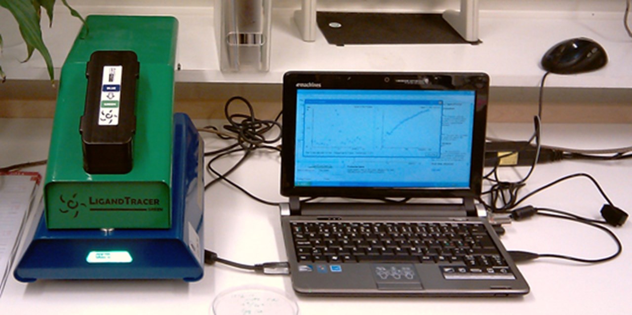
LigandTracer® Green – an instrument for real-time quantification of binding of fluorescently labeled biomolecules or bacterial cells to immobilized cells or receptors.
LigandTracer® Green offers a simple and accurate method for detection and quantification of biomolecular/cells interactions in real‐time using fluorescently labeled biomolecules. Measurements of e.g. on‐off rate, affinity or retention can be made using blue (~488nm) and green (~535nm) fluorophores, e.g. FITC, Alexa Fluor® 488, as labels. A detector for Yellow (590nm) - Red (632nm) for Rhodamine Red and similar fluorophores is also available.
Although the instrument has been originally developed for monitoring of ligands (proteins including antibodies) interactions with surface receptors of immobilized cells, this technology has also been demonstrated to be applicable for protein to protein, protein to tissue or bacterial cells to immobilized receptors (Fig. B) or to cells interactions.
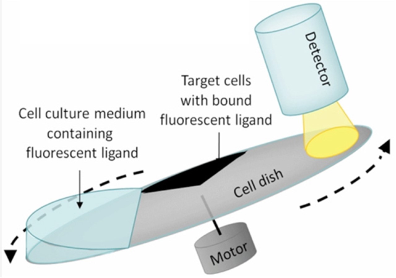
Fig.A: The principle of rotating RIA with LigandTracer Green (the material is from an application note at LigandTracer homepage, www.ligandtracer.com).
The LigandTracer technology is based on 'a rotating RIA' (Radio Immuno Assay): kinetic data is generated by repeatedly measuring bound ligand concentration in a cell- (alt. receptor protein-) covered 'target' area of a circular dish (Fig. A). As a dish is rotating in a slightly inclined position, the target area is consequently passes liquid, containing fluorescently labeled ligand, and an essentially liquid-free area under a detector mounted over the elevated part of a dish. As the ligand binds to the receptors, an intensity of the detected signal will increase. The differential signal (when a signal from the reference area is subtracted from a signal of the target area) represents a background corrected measure of the amount of ligand bound to the immobilized cells or receptors (Fig. B).
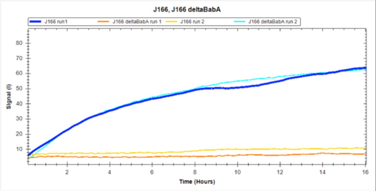
Fig. B: An example of our assay showing binding kinetics of FITC-labeled H. pylori J166 strain to a receptor (HSA-Leb) - coated area of a dish. The signal is a background corrected (coated area minus reference area coated with HSA-Lea) and is a measure of the amount of bacterial cells attached to immobilized Leb receptors. The negative control is a J166 deltaBabA mutant showing no specific binding to Leb. Binding of each strain is monitored in two independent runs.
The instrument is produced by Ridgeview Instruments AB, Uppsala, Sweden
Owned/Located: Umeå University, Department of Medical Biochemistry and Biophysics, group Thomas Borén.
The information about LigantTracer and underlying technology is based on the materials provided at www.ligandtracer.com. Please contact this site for more detailed information, references and application notes.