"False"
Skip to content
printicon
Main menu hidden.

Image: Mattias Pettersson

Published: 2022-11-30

Advanced microscope provides frozen snapshots at atomic level

FEATURE In the cryo-electron microscopes at Umeå University, cells, proteins and viruses are studied in detail – after first beeing frozen to minus 190 degrees Celsius. The state-of-the-art equipment takes pictures of events that were previously impossible to capture.

Text: Sara-Lena Brännström

A four metre high device for looking at nature's smallest building blocks. That's one way to describe the large cryo-electron microscope, which has opened up enormous possibilities for researchers – not just in Umeå but throughout the country.   
  
The technology is highly advanced and was awarded the Nobel Prize in Chemistry in 2017. Using cryo-electron microscopy, scientists in Umeå have recently been able to see, among other things, how polioviruses take over human cells, high-resolution images of details of photosynthesis and how misfolded proteins cause the so-called Skellefteå disease.

It means a lot for Swedish researchers to have access to the absolute cutting edge of equipment

“We can see what a protein looks like when it's working properly in a healthy cell and what a protein looks like when it causes disease, and compare the structures. Or how a virus infects a cell, the coronavirus for example. Understanding how the virus affects the cell's proteins can lead to the development of new drugs,” says Linda Sandblad, researcher and Director of the Umeå Centre for Electron Microscopy.  

Well-controlled temperature

The equipment is important for research groups in fields ranging from biochemistry to cell biology in medicine and plant biology.  
  
“It means a lot for Swedish researchers to have access to the absolute cutting edge of equipment. That makes Swedish research competitive in the world,” she says.  

The large Titan Krios cryo-electron microscope has been at the university since 2016 and is located in a very well-controlled environment in the basement of the KBC building. Humidity, temperature and vibrations are carefully regulated. The instrument works with an enormous precision.  
  
“It has a magnification and resolution that makes it possible to see proteins at angstrom level, that is to say individual atoms, when processing the image. That is the benefit for molecular biology, to be able to see how proteins are shaped and the positions of the atoms in the proteins,” says Linda Sandblad.

Shows three-dimensional image

What is unique about cryo-electron microscopy is that the material to be studied is cooled down to minus 190 degrees Celsius. The sample is placed in the microscope, which takes many thousands of images from different angles. These are then put together to form a highly detailed three-dimensional image.

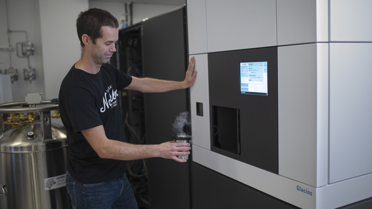
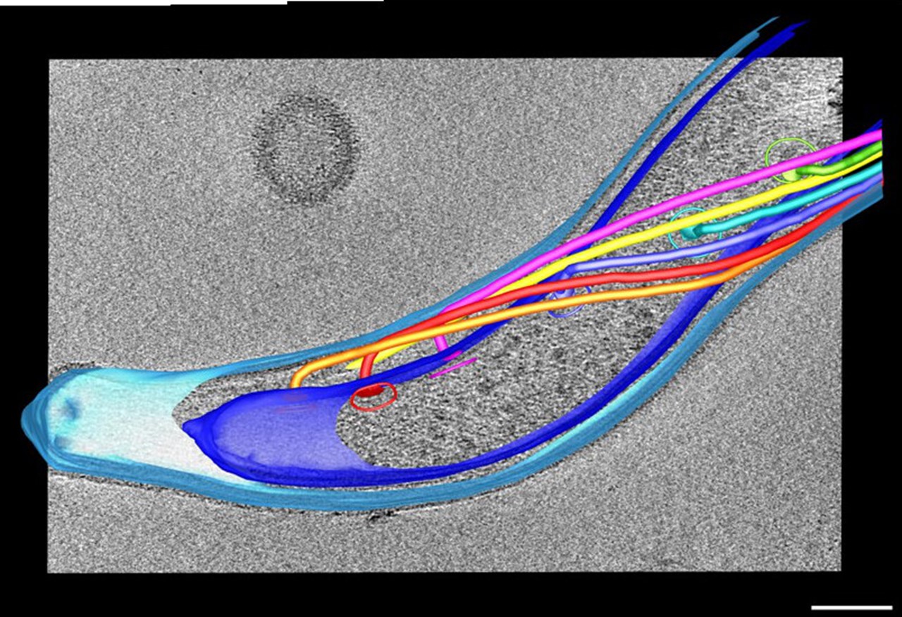
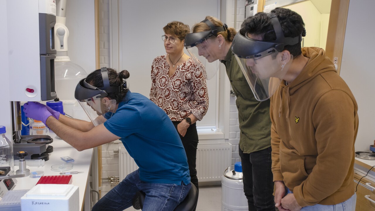
FACTS How cryoelectron microscopy works

The method involves rapidly freezing biomolecules and thus capturing them in mid-motion.

First, a very thin film of the biological material to be studied is created on a fine-mesh metal grid. The sample is then flash-frozen to minus 190 degrees and placed in the cryo-electron microscope. An electron gun shoots electrons that travel through a magnetic column, producing the magnification.

At the other end of the microscope is a detector that reads the electrons that have passed through the frozen biological sample, and saves the image. Image processing requires powerful computers and makes it possible to display 3D images of the biological material.

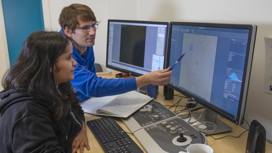
Pictured: Kasturika Shankar and Jan Silhán are starting a data collection at Glacios.

Only two universities in Sweden offer the use of cryo-electron microscopes to researchers nationwide: Umeå and Stockholm. In spring 2021, the two universities together received a grant of SEK 30 million from the Swedish Research Council, the Kempe Foundation and SciLifeLab to upgrade the equipment.  
  
As a result, the microscope in Umeå got a "little sister": Glacios. Similar to its older sibling, but smaller, it is used as a first station, where researchers can examine the quality of samples before they are passed on to the larger microscope.   

Shorter waiting times

Glacios is in operation all the time, around the clock. Many research groups use it in their studies and it is also used for training.  
  
“It gives us much greater capacity so that we can deliver more image data for structural and cell biology to our researchers in a short time. Researchers should not have to wait several months, they should be able to come and do experiments when they need to,” says Linda Sandblad.  

In the large cryo-electron microscope, a new electron detector has recently been installed, the very latest on the market. It is still being fine-tuned and will soon be able to take even more refined images, even faster.

Right now, cryo-electron microscopes are playing a crucial role in a variety of research projects. Having them in Umeå means a lot to the university, says Linda Sandblad.

“It means that we can provide PhD students and postdocs with technical expertise that is in high demand. It also makes Umeå University attractive to researchers. Students and researchers from all over the world come to Umeå because we have the cryo-electron microscopes,” she says.

Read more about Umeå Centre for Elecron Microscopy (UCEM)