"False"
Skip to content
printicon
Main menu hidden.

Light Microscopy

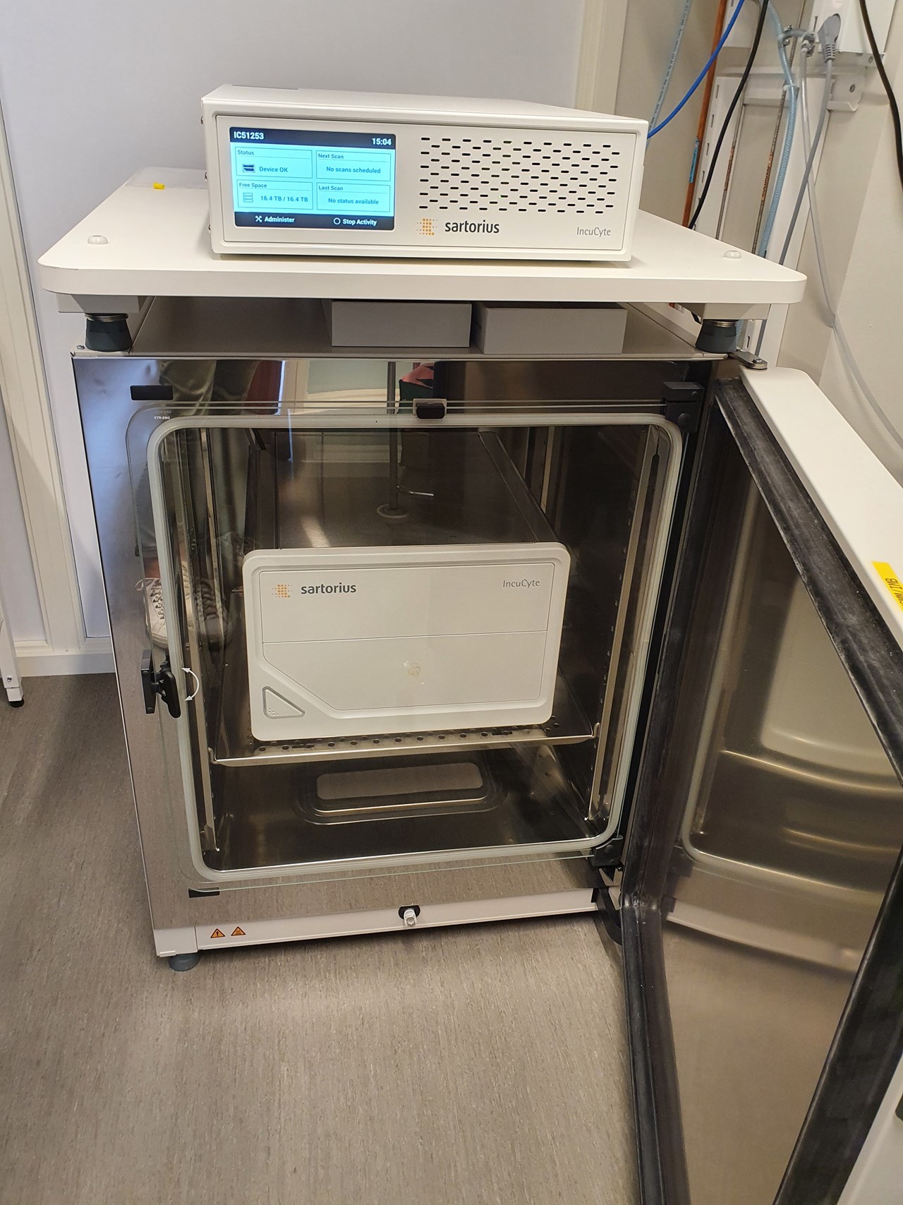
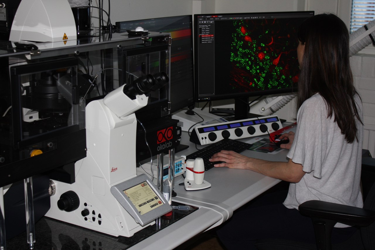
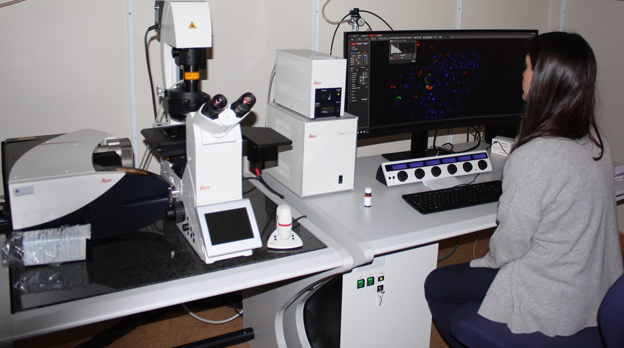
Within light microscopy we provide advanced live cell imaging techniques including FLIM, FRAP, FLIM-FRET, STORM, TIRF, Spinning disk confocal and 4D confocal.

Contact:

Facility Director

Facility Manager

Light Microscopy Booking:

To book any of our instruments, please visit our Online Booking System

Note: The e-booking requires that you have had previous education on the specific system. If you dont have access to e-booking or if you want education or support please contact Irene Martinez

Rules:

  • To operate the instruments on your own, you must have a licence. The licence will allow you to access the e-booking system and the server
  • Data should be transferred to the server and to your personal storage. You cannot store your images in the server or the microscope computer.
  • For funding purposes, it is essential to acknowledge BICU in all publications that include data derived from the facility. Include this statement: "Microscopy was performed at the Biochemical Imaging Centre Umeå.

Light Microscopy Instruments and Workstations

Leica SP8 FALCON Confocal

More Details

  • Location:
    Chemistry Department 4th floor corridor A4
  • Room number:
    A4.41.01
  • Price:
    250kr/hr - Driver's license 2000kr
  • Objectives:
    • HC PL FLUOTAR 10x/0.30
    • HC PL APO 20x/0.75 CS2
    • HC PL APO 63x/1.20 Water motCORR CS2
    • HC PL APO 63x/1.40 OIL CS2
  • Laser Lines:
    • Diode 405nm
    • Argon Laser (458nm, 476nm, 488nm, 496nm and 514nm)
    • WLL (White light laser). Tunable excitation from 470 to 670nm in steps of 1nm
  • Techniques:
    • Confocal Imaging
    • FLIM (biosensing, species separation, FLIM-FRET)
    • FRAP
  • Other specifications:
    • One PMT detector, two HyD SP GaAsP detectors and one HyD SP generation2 detector optimized for FLIM
    • Motorized Focus with AFC
    • Resonant Scanner 12 kHz
    • Live cell incubation
    • Microinjector
       

Leica SP8 Confocal

More Details

  • Location:
    Molecular Biology
  • Room number:
    6L-021
  • Price:
    250kr/hr - Driver's license 2000kr
  • Objectives:
    • HC PL APO 20x/0.75 IMM CORR CS2
    • HC PL APO 40x/1.30 Oil CS2
    • HC PL APO 63x/1.40 OIL CS2
  • Laser Lines:
    Diode 405nm, Diode 638nm, OPSL 488nm, OPSL 552nm
  • Techniques:
    • Confocal Imaging
    • Resonant Scanner 8 kHz

Zeiss Spinning Disk Confocal

More Details

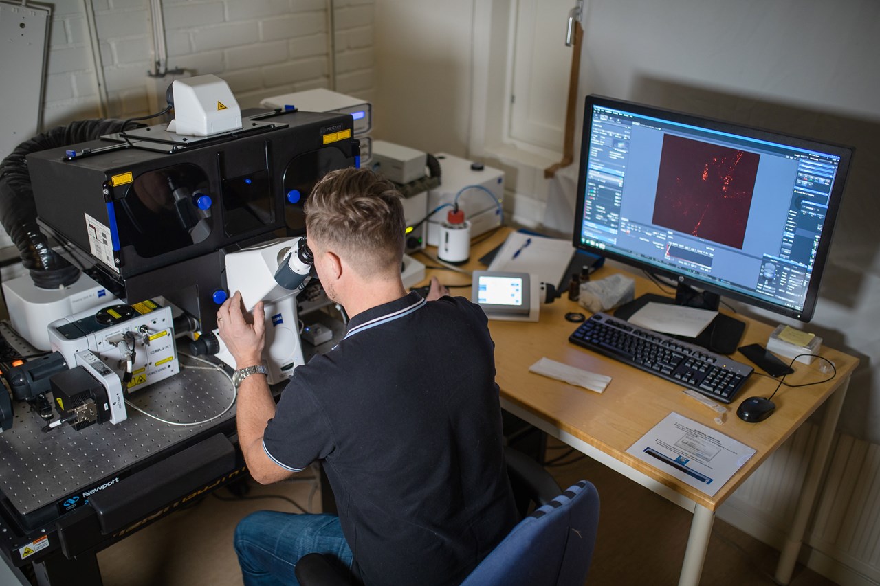
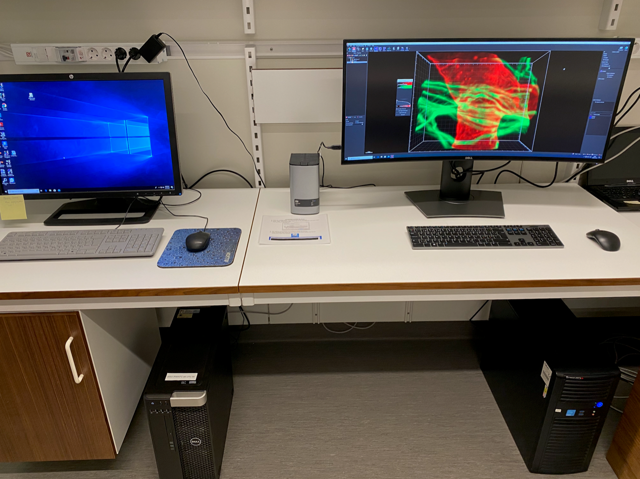
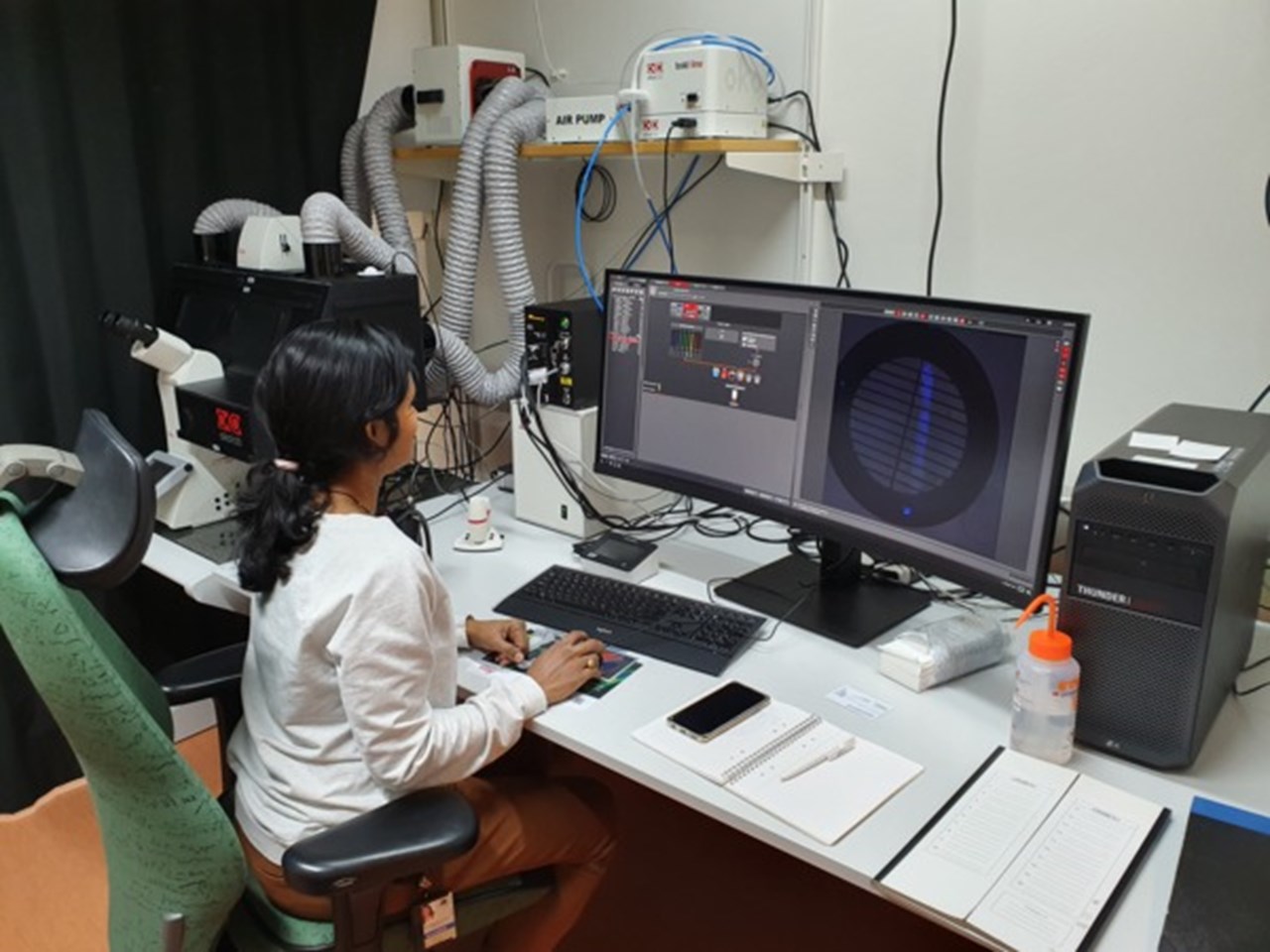
Zeiss Cell Observer Spinning Disk Confocal, equipped with two EMCCD cameras to perform confocal and TIRF microscopy.

  • Location:
    IMB: Department of IntegrativeMedical Biology. 4thfloor
  • Room:
    C4 15 17
  • Price:
    250kr/hr - Driver's license 2000kr
  • Objectives:
    20X (NA 0.8); 40X W (NA 1.2); 63X oil (NA 1.40) DIC; 100X Tirf oil (NA 1.46) DIC
  • Laser lines:
    405nm, 488nm, 561nm and 647nm
  • Techniques:
    • Confocal imaging (1000 frames/sec)
    • TIRF microscopy
  • Other specifications:
    • Live cell incubationDefinite Focus system
    • Direct FRAP
    • Microfuidics
  • Manual for Download:

Zeiss LSM 710 Confocal

More Details

  • Location:
    Oncology Department. Hospital Building 6M, 3rd floor
  • Price:
    250kr/hr - Driver's license 2000kr
  • Objectives:
    • Fluar 5x/0.25
    • Plan Apochromat 10x/0.45
    • Plan Apochromat 20x/0.8
    • C-Apochromat 40x/1.2 W
    • Plan Apochromat 63x/1.4 Oil DIC III
  • Laser Lines:
    405nm, Argon Laser (458nm, 488nm, 514nm), 561nm and 633nm
  • Techniques:
    Confocal Imaging

Nikon A1R Confocal

More Details

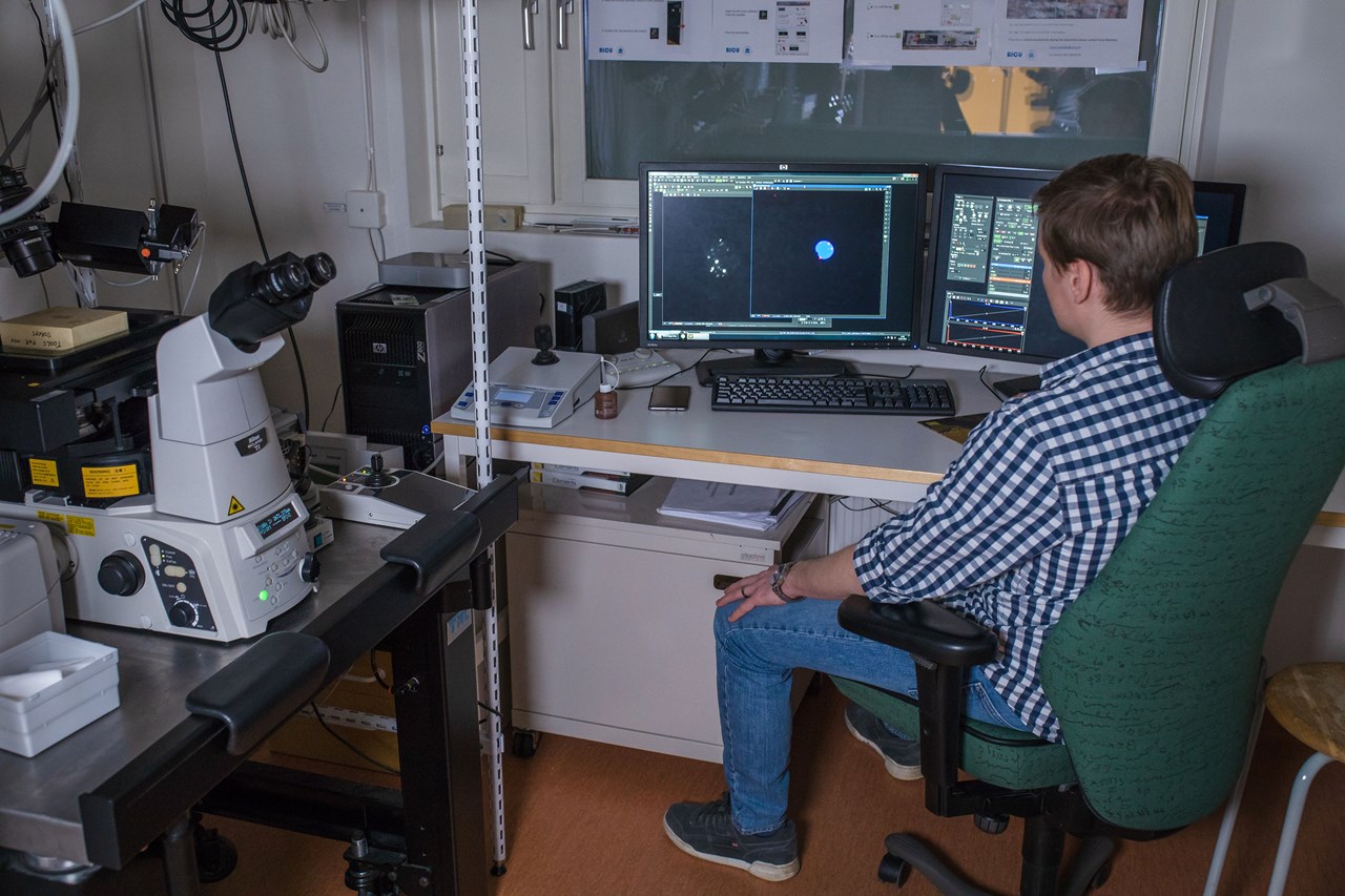
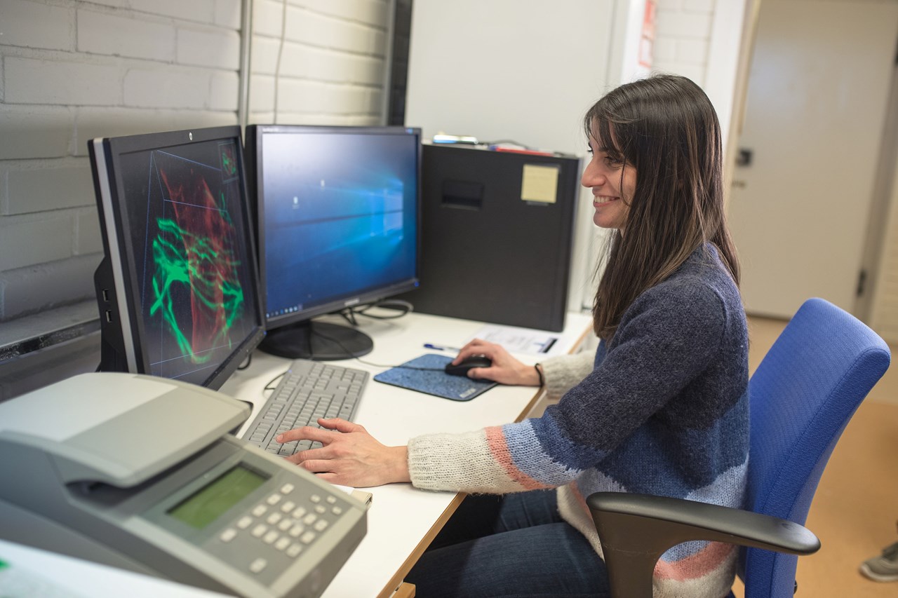
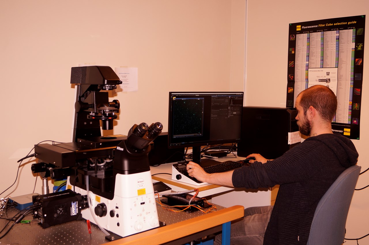
Nikon A1R confocal (LSM) controlled by Nikon NIS Elements interface with a Nikon Eclipse Ti-E inverted microscope.

  • Location:
    KBC Building 6thfloor Department of Medical Biochemistry and Biophysics.
  • Room:
    A6 44 15
  • Price:
    250kr/hr - Driver's license 2000kr
  • Objectives:
    10X (NA 0.45) DIC 20X (NA 0.75) DIC 60X oil (NA 1.40) DIC 100X Tirf oil (NA 1.49) DIC
  • Laser lines:
    405nm, 457, 477, 488, 514nm, 561nm and 647nm
  • Techniques:
    • Confocal microscopy (30 frames/sec)
    • Full spectral resolution (spectral and conventional detectors)
    • STORM microscopy
    • TIRF microscopy
    • Epifluoresence microscopy
  • Other specifications:
    • Live cell incubation
    • Micromanipulators
    • Perfect focus system
  • Manuals for Download:


Leica Widefield Thunder

More Details

  • Location:
    KBC Building 6th floor Department of Medical Biochemistry and Biophysics.
  • Room:
    A6 44 15
  • Price:
    150kr/hr - Driver's License 2000kr
  • Objectives:
    • N PLAN 5x/0.12 PH0
    • HC PL FLUOT AR 10x/0.32 PH1
    • HC PL FL L 20x/0.40 CORR PH1
    • HC PL APO 20x/0.80
    • HC PL FLUOTAR L 40x/0.60 CORR
    • HC PL APO 40x/0.95 CORR
    • HC PL APO 63x/1.40-0.60 OIL
    • HC PL FLUOTAR 100x/1.32 OIL PH3
  • Techniques:
    • Widefield
    • Live cell imaging
    • Computational clearance: Thunder

Nikon Widefield/Tirf

More Details

  • Location:
    NUS, building 6M
  • Room:
    06M-RUM-6098
  • Price:
    • Wide field: 150kr per hour. Driver’s license: 2000kr
    • TIRF unit: 200kr per hour. Driver’s licence: 2000kr
    • FRAP unit: 200kr per hour. Driver’s licence: 2000kr
  • Objectives:
    • Plan Apo λ 4x/0.20
    • Plan Apo λ 20x/0.75
    • S Plan Fluor RLWD 40x/0.60
    • Apo TIRF 60x/1.49 Oil
  • Illumination:
    • LED (390, 475, 510, 555, 575, and 635)
    • Lasers (Tirf microscopy) 488nm (50mW) and 561nm (50mW)
  • Filter cubes currently installed:
    • DAPI Filter Cube (32mm) (EX 392/23, DM 409, BA 447/60)
    • FITC Filter Cube (32mm) (EX 474/27, DM 495, BA 525/45)
    • mCherry Filter Cube (32mm) (EX 562/40, DM 593, BA 640/75)
    • Cy5 Filter Cube (32mm) (EX 635/18, DM 652, BA 680/42)
    • TIRF-Quad Filterset 405/488/561/640 Nikon
  • Techniques:
    • Widefield
    • Live cell imaging
    • Tirf microscopy
  • Other specifications:
    • Live cell incubation
  • Supporting Documents:

IncuCyte Live-cell Analysis System

More Details

  • Location:
    IMB, D2 wing 5th floor
  • Room:
    D2-524
  • Price:
    20kr/hr
  • Objectives:
    10x, 20x
  • Techniques:
    Live cell imaging: Angiogenesis, Antibody internalization, Apoptosis, Cell cycle, Chemotaxis, Cytotoxicity, Immune cell killing and clustering, Immunocytochemistry, NETosis, Neurite analysis, Phagocytosis, Proliferation, Scratch wound migration and invasion, Spheroids.
  • More information:
    https://www.essenbioscience.com/en/products/incucyte/incucyte-s3/

Mass Photometer

More Details

Imaris Workstation

More Details

  • Location:
    IMB, D2 wing 5th floor
  • Room:
    D2-524
Latest update: 2023-12-14