
NEWS PhD student from University of Helsinki collaborates with UCEM to identify a novel player of peroxisomal biogenesis.
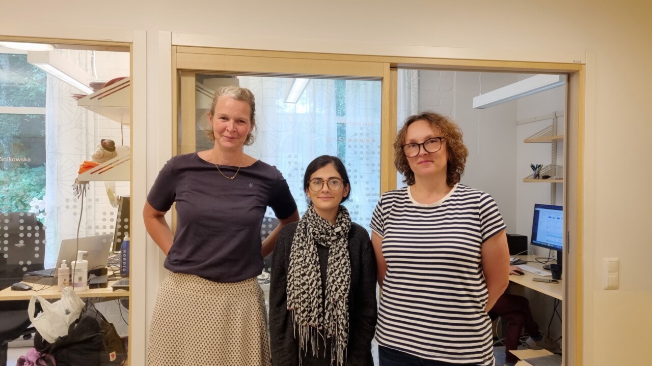
Collaborative project between UCEM and University of Helsinki (from left to right): Linda Sandblad (head of UCEM), Kaneez Fatima (PhD student from University of Helsinki) and Agnieszka Ziolkowska (personnel at UCEM
ImageKaneez FatimaThe Umeå Centre for Electron Microscopy (UCEM) at Umeå University has started a collaborative project with Kaneez Fatima, a third year PhD student from the Institute of Biotechnology, University of Helsinki, and Helsinki Institute of Life Science (HiLIFE). Fatima’s area of expertise lies in the field of cell and molecular biology, with a specific focus on peroxisomal biogenesis. This collaboration can provide the final missing piece of the puzzle addressing her research question on peroxisomal biogenesis.
Peroxisomes are challenging to be identified with electron microscopy because of their resemblance with lipid droplets and other organelles. They are very small as well, and they are “shapeshifters”, meaning that they change their shape and size according to the cell conditions present. They also exist in both premature and mature forms. Fatima’s group is using mammalian cell lines and they have already successfully identified some novel proteins of the peroxisomal biogenesis. Together with UCEM, Fatima is working on characterizing one of these proteins, an endoplasmic reticulum protein, which has shown to have a great impact on peroxisomal maintenance.
“There hasn’t been a systematic approach used to study peroxisomal biogenesis and how proteins reach peroxisomes remains unclear. We know few of the membrane proteins involved and some additional proteins. High-resolution imaging of peroxisomes is an essential step in our study to reveal mechanisms of peroxisomal biogenesis. However, it is a very challenging task. If we manage to do so here with the CLEM technique, that will be a big breakthrough, and UCEM has the right expertise to achieve this”, says Kaneez Fatima.
She visited Umeå first-time last year to participate at the Advanced Correlative Light and Electron Microscopy (CLEM) workshop, organized by UCEM and The Laboratory for Molecular Infection Medicine Sweden (MIMS), where she realized that this technique is exactly what she needs for her project. At UCEM, Naga Venkata Gayathri Vegesna, Head of CLEM unit, Facility manager, and Agnieszka Ziolkowska, Facility manager and expert in TEM, are collaborating with her.
“We had the first project meeting with Fatima right after the CLEM workshop, and we decided to join forces since we have the expertise here what she needed for her project. After some follow up meetings and further experiments, we determined that the samples need to be fixed and sectioned under cryo conditions, then we use the Tokuyasu method to access the epitopes and antigens for both immunogold labelling and fluorescence imaging, which then we can correlate with each other”, explains Gayathri Vegesna the methodology to be used.
For the CLEM method, the necessary infrastructure is provided by the combination of Umeå Centre for Electron Microscopy and Biochemical Imaging Centre Umeå (BICU).
Within a two-week period, they see the results and can start the analysis of the images. Mapping the mechanism of peroxisomal biogenesis would help to better understand pathological conditions associated with peroxisomal dysfunction including neurological and metabolic diseases and certain types of cancer.
The supervisor of Kaneez Fatima is Dr Svetlana Konovalova, and co-supervisor Prof Pekka Katajisto.