
NEWS By examining the cornea of the eye with a special microscope it may be possible within ten minutes to diagnose if a person with type 2 diabetes has nerve damage. This according to a study among diabetics in Skellefteå, north Sweden.
“Although there is currently no cure, it’s always an advantage to detect changes in the nerves early. Therefore, it’s valuable to find a fast and safe diagnostic method,” says Professor Olov Rolandsson, at the Department of Public Health and Clinical Medicine, Family Medicine, at Umeå University, who was responsible for the study.
Type 2 diabetes can lead to impaired nerve function, half of those who have had type 2 diabetes for more than ten years have a decrease in nerve functions, which usually begins in their feet. The decrease may be expressed as reduced sensibility or pain and increases the risk of wounds. Sometimes the reduction can also cause the need for an amputation. There is currently no cure for this type of nerve damage, and the methods available so far at clinics to diagnose the condition are not entirely optimal.
 When type 2 diabetes develops, small nerve fibres in the peripheral nervous system begin losing their function even before symptoms become apparent. These small peripheral nerve fibres are found in the skin, but also in the cornea of the eye. Because the cornea is transparent, it is an ideal place to study nerve damage that occurs in diabetics.
In the study with 82 people from Skellefteå, with or without type 2 diabetes, the participants' eyes were examined with a special microscope. The study found that the corneal nerve density was lower in individuals with type 2 diabetes compared to healthy subjects. If the individual had had type 2 diabetes for a longer period of time, nerve density was even more reduced.
The study is the first of its kind in Sweden. Through cooperation between a Swedish research team and researchers in Germany, Italy and Norway, a method has been developed to create images in three dimensions of corneal nerves. The examination only takes ten minutes and does not cause any discomfort to the patients. The research team has also developed an automated analysis programme that allows the method to be used in general health care. Microscopic examination of the cornea occurs internationally, but the Skellefteå study is the first in the world to use the entire new method for assessing the degree of nerve damage in diabetics.
“There are good conditions for this new diagnostic method to be widely  introduced in health care,” says Olov Rolandsson.
Olov Rolandsson at Umeå University in Sweden led the Skellefteå study, while the technical development and microscopy survey was conducted by Docent Neil Lagali at Linköping University. The study is published in the scientific journal IOVS, Investigative Ophtalmology & Visual Science.
Olov RolandssonProfessorDepartment of Public Health and Clinical MedicineUmeå UniversityPhone: +46 90 785 35 71Mobile: +46 70 590 20 52
E-mail: olov.rolandsson@umu.se
Press portrait photo on Olov Rolandsson for free download. Photo: Mattias Pettersson.
Reduced Corneal Nerve Fiber Density in Type 2 Diabetes at Wide-Area Mosaic AnalysisIOVS, Investigative Ophtalmology & Visual ScienceLagali NS, Allgeier S, Guimarães P, Badian RA, Ruggeri A, Köhler B, Utter TP, Peebo B, Peterson M, Dahlin LB, Rolandsson O. Reduced Corneal Nerve Fiber Density in Type 2 Diabetes at Wide-Area Mosaic Analysis. Invest Ophthalmol Vis Sci. 2017 Dec 1; 58 (14): 6318-6327. doi: 10.1167 / iovs.17-22257.
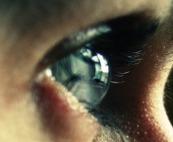
Photo eye: Mostphotos
Editor: Ola Nilsson