

New microscope will reveal 3D cell structures at nanoscales
Umeå Centre for Electron Microscopy (UCEM) recently inaugurated an advanced microscope which will contribute greatly to research in several life science fields. The new instrument is a FIB-SEM (Focused Ion Beam – Scanning Electron Microscope), specifically designed to reveal thin layers of cells whilst operating at extremely low temperatures, enabling detailed studies of biological samples at the nanoscale.
Published: 2024-11-29 Updated: 2024-12-04, 09:33
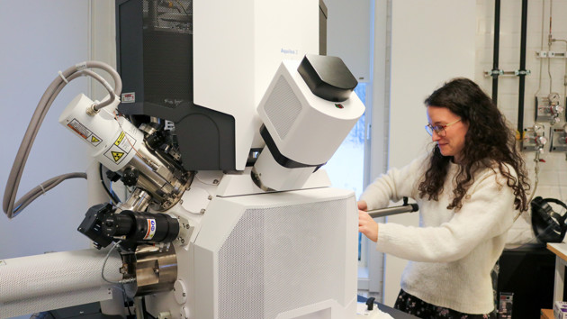
Erin Schexnaydre, staff scientists at UCEM, shows the new FIB-SEM instrument "Aquilos 2". The microscope can image biological samples at nanoscales, whilst the sample is kept at freezin -180 degrees Celsius.
ImageAnna ShevtsovaOperating at -180°C
Here in Umeå, we are used to freezing conditions, but in the labs at Umeå Centre for Electron Microscopy (UCEM), they take the cold one step further.
“Having cells frozen to very low temperatures preserve the biological structures within them, such as proteins, and allows us to study the functions of these proteins”, explains Erin Schexnaydre, staff scientist at UCEM, who will be the manager of the new instrument, named Aquilos 2.
Before the microscope is put to use, samples are frozen to below -180 °C. This is done in a rapid cooling process, preventing ice crystals from forming in the sample which would otherwise cause damage to the sensitive biological structures.
“However, the sample is at this stage too thick to be directly imaged by an electron microscope," says Erin Schexnaydre, “which is why we need the combination of both the focused ion beam and the electron microscope.”
By using a beam of charged particles – ions, Aquilos 2 can create thin layers of frozen samples – called cryo-lamella. Cryo-lamella can be as thin as 200 nanometers, which is 500 times thinner than human hair, and much thinner than the average size of a human cell. This technique opens the cell for detailed studies of its inner structures in 3D and is ideal for obtaining detailed images of the interior of cells.
Round-the-clock science
Although purchase was finalised already at the end of last year, it has been a long wait for the new microscope to be up and running. Following some necessary room reconfigurations, the instrument is now in use and was celebrated with a cheerful inauguration at UCEM and a contest of the best electron microscope image.

Cheerful mood at UCEM as the new FIB-SEM microscope "Aquilos 2" was inaugurated in November. It was celebrated with drinks and a competition of the best electron microscopy image.
ImageAnna Shevtsova“Aquilos 2 is designed to use the ion-beam overnight, which increases the throughput of cryo-lamella production and thus the collection of data,” says Erin Schexnaydre. Obtaining more data quicker allows the researchers to explore complex biological structures more efficiently, opening doors to new scientific discoveries.
Who has access to the new microscope? “Anyone at Umeå University, or collaborators in SciLifeLab, who have projects requiring this microscope can be trained by the UCEM staff to use the new instrument,” says Erin Schexnaydre, who hopes it will come in good use for many exciting research projects.
What is a Scanning Electron Microscope?
A scanning electron microscope (SEM) is a type of electron microscope that produces images of a sample by scanning the surface with a focused beam of electrons. The electrons interact with atoms in the sample, producing various signals that contain information about the structure of the surface and composition of the sample.
Image: Madeleine Ramstedt

Kontakt för mer information


Umeå Centre for Electron Microscopy (UCEM)
UCEM is a joint resource for research and higher education in electron microscopy techniques.

SciLifeLab Site Umeå
SciLifeLab national site at Umeå (UMU and SLU) is a local site of the national SciLifeLab network.

Umeå Centre for Microbial Research UCMR
UCMR is a Linnaeus Centre of Excellence promoting cutting-edge microbial research at Umeå University.
Nordic EMBL Partnership for Molecular Medicine




Supporting organisations


