
About the scientific article:
Jönsson, M., et al: Engineered calcium-regulated affinity protein for efficient internalization and lysosomal toxin delivery. PNAS. 10.1073/pnas.2509081122
NEWS Cancer-fighting antibody drugs are designed to penetrate tumor cells and release a lethal payload deep within, but too often they don’t make it that far. A new study shows how this Trojan Horse strategy works better by exploiting calcium differences outside and inside cells.
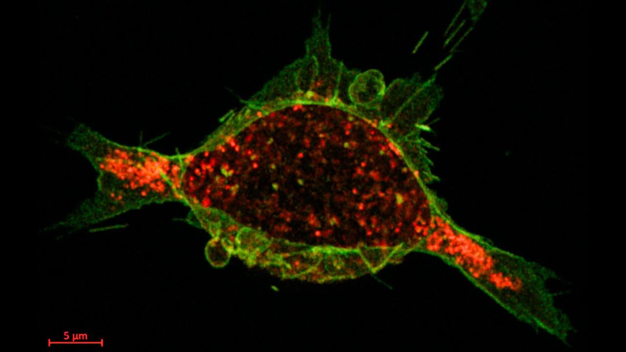
The calcium-regulated protein drug (green) and tumor cell receptors (red) have bonded and internalization is underway, 40 seconds after administration.
ImageKTHThe calcium switch is built into the drug design
A research team from KTH, Stanford University and Umeå University has developed a calcium activated delivery system that could enable more precise treatment, with lower doses and less collateral damage to healthy tissue. The results have been published in PNAS, the journal of the National Academy of Sciences.
The concept takes aim at a common challenge with targeted drugs, which tend to cling too tightly to receptors expressed by tumors. On the positive side, that strong bond blocks receptors’ tumor growth signals. But ADCs (Antibody–Drug Conjugates) are also meant to attack and kill, and too often the protein can become stuck without ever penetrating deeper into the cell’s real intended goal: an acidic compartment called the lysosome. There, in the kill zone, the targeting protein can be broken down, thus unleashing toxin that causes cell death.
To avoid that problem, the researchers developed a calcium-sensitive switch that binds strongly to the cancer cell receptor on the outside of the cell where relatively high calcium concentrations are found, in the blood and the extracellular fluid.
Once bound together, the drug-loaded protein (or calcium-regulated affinity, CaRA) and epidermal growth factor receptor (EGFR) are pulled inside the cell, into compartments with gradually lower levels of calcium. And because their bond is calcium dependent, the receptor and CaRA eventually go their separate ways: the receptor can recycle back to the membrane, while CaRA continues carrying its payload toward the lysosome.
“The calcium switch is built into the drug design. It senses calcium levels and changes its grip automatically,” says Sophia Hober, professor at KTH Royal Institute of Technology who led the study.

From Umeå University, Professor Magnus Wolf-Watz's group participated in the study published in PNAS.
ImageMattias PetterssonThe study was performed on living human cancer cell lines, using a payload of the cytotoxin, mertansine DM1. The drug conjugate showed a very high potency and it is highly selective—it only killed cells that overexpress EGFR, leaving healthy or low-EGFR cells unharmed. The researchers emphasize that this shows specific targeting and a strong therapeutic window, which is critical for reducing side effects.
Leon Schierholz, a doctoral student in Magnus Wolf-Watz's research group at Umeå University, has determined a low-resolution structure of the complex between CaRA; and the extracellular domain of the EGFR receptor. The structure has been determined to a resolution of approximately 6 Å using the single particle cryoEM technique on data collected at the Umeå Centre for Electron Microscopy, UCEM, at Umeå University.
The structure provides a fundamental molecular understanding of the high affinity of the complex.

Doctoral student Léon Schierholz has spent many hours at the large microscope at the UCEM technology platform at Umeå University.
ImageHans Karlsson“We are now moving forward and aiming to come up with a high-resolution structure with a resolution below 3.5 Å that can allow us to make an atomic model that can be used to further improve the properties of CaRA. The data is of very good quality for this relatively small complex,” says Leon Schierholz, who in this context wants to put the spotlight on his colleague Max Renner, who is highly involved in completing the atomic model.
Jönsson, M., et al: Engineered calcium-regulated affinity protein for efficient internalization and lysosomal toxin delivery. PNAS. 10.1073/pnas.2509081122
This is a technique used in structural biology to determine the structure of biomolecules such as proteins at near-atomic resolution. The method involves collecting a large number of 2D images of the same molecule in different orientations from frozen samples and then combining them to create a 3D model of the molecule.

