
About the scientific article:
Sengupta, P., Gillet, N., Obi, I. and Sabouri, N.: Mechanistic insights into PCBP1-driven unfolding of selected i-motif DNA at G1/S checkpoint. Nature Communications. 2026. DOI: 10.1038/s41467-026-68822-5
NEWS DNA’s iconic double helix does more than “just” store genetic information. Under certain conditions it can temporarily fold into unusual shapes. Researchers at Umeå University have now shown that one such structure, known as i-DNA, not only forms in living cells but also acts as a regulatory bottleneck linked to cancer.

Postdoctoral researcher Pallabi Sengupta studies i-DNA, a field of research that is still in its early stages.
ImageMattias Petterssona kind of ‘peek‑a‑boo structure’ in the DNA molecule
“You can think of i-DNA as a kind of ‘peek‑a‑boo structure’ in the DNA molecule. Its formation is tightly controlled in time and it must be resolved at precisely the right moment. We believe it plays an important role in gene regulation, because these structures can appear and disappear in sync with changes in the cell’s state,” says first author Pallabi Sengupta, postdoctoral researcher at the Department of Medical Biochemistry and Biophysics at Umeå University. The study is now published in Nature Communications.
The familiar double helix can be imagined as a twisted ladder with sugar‑phosphate backbones as side rails and base pairs – adenine (A) paired with thymine (T), and cytosine (C) paired with guanine (G) – forming the rungs.
i-DNA, however, bears little resemblance to this shape. Instead, it is more like a distorted, self‑folded ladder tied into a knot. It consists of a single DNA strand folding back on itself to form a four‑stranded structure. At the molecular level, the structure is held together not by standard A–T and C–G base pairs, but by pairs of cytosines.
These rare, short‑lived structures appear and disappear depending on the cellular environment. For decades, they were dismissed as too unstable to exist inside cells and regarded as laboratory artifacts. With new experimental techniques, researchers in Umeå can now demonstrate that i-DNA does form, but only briefly, just before DNA replication begins.
The study further shows that the protein PCBP1 acts as a critical regulator. It unwinds i-DNA at the right moment, allowing the DNA replication machinery to proceed. If the structures fail to open in time, they block replication, increasing the risk of DNA damage – a hallmark of heightened cancer vulnerability.
The researchers also discovered that i-DNA is not uniform: some structures are easy to unwind, while others are highly resistant, depending on the underlying DNA sequence.
“The more cytosine base pairs that hold the knot together, the harder it is to resolve. In some cases, hybrid structures can form, making i-DNA even more stable,” explains Nasim Sabouri, professor at the Department of Medical Biochemistry and Biophysics at Umeå University, who led the study.
Notably, many i-DNA structures are located in regulatory regions of oncogenes – genes that drive cancer development – suggesting a direct link between i-DNA and disease.
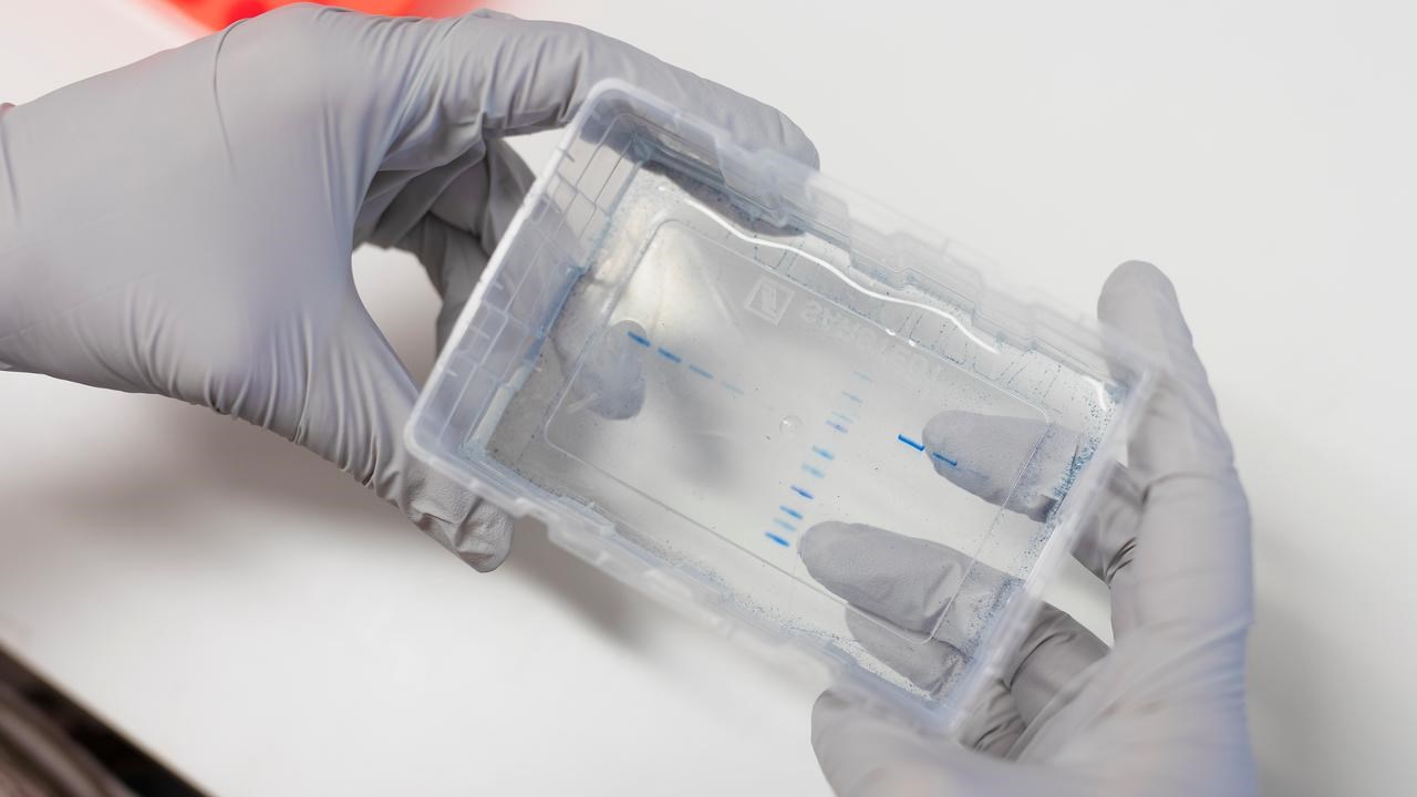
The image shows how proteins become visible in a gel after being isolated from cells.
ImageMattias PetterssonTo study these short-lived structures, the team combined biochemical assays, computational modelling and cell biology. They successfully visualized how PCBP1 progressively opens i-DNA and captured the structures in living cells at the exact moment in the cell cycle when they appear.
“By connecting molecular mechanisms to actual effects in cells, we can show that this is biologically relevant and not a laboratory phenomenon,” says Ikenna Obi, staff scientist at the Department of Medical Biochemistry and Biophysics at Umeå University.
The discovery reframes i-DNA from a molecular oddity to a potential weakness in cancer cells. Because cancer cells often experience high replication stress attempting to divide so rapidly that their DNA replication machinery approaches breakdown, any disruption in i-DNA handling may have severe consequences.
“If we can influence i-DNA or the protein that unwinds it, we may be able to push cancer cells beyond their tolerance limit. This opens completely new avenues for drug development,” says Nasim Sabouri.
The study was conducted in collaboration with Natacha Gillet, researcher at the Centre National de la Recherche Scientifique (CNRS) in France. It was funded by Cancerfonden, the Wenner-Gren Foundations, and the Knut and Alice Wallenberg Foundation.
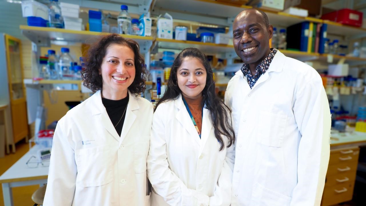
Nasim Sabouri, Pallabi Sengupta, and Ikenna Obi. The team will now investigate which cellular conditions promote i-DNA formation and whether these short‑lived structures can serve as new targets for diagnostics and cancer therapy.
ImageRebecca ForsbergSengupta, P., Gillet, N., Obi, I. and Sabouri, N.: Mechanistic insights into PCBP1-driven unfolding of selected i-motif DNA at G1/S checkpoint. Nature Communications. 2026. DOI: 10.1038/s41467-026-68822-5

