
NEWS With her many years of experience, Senior Research Engineer Fouzia Bano is instrumental in making atomic force microscope accessible to other researchers both within the Department of Clinical Microbiology and to other research communities at Umeå University.
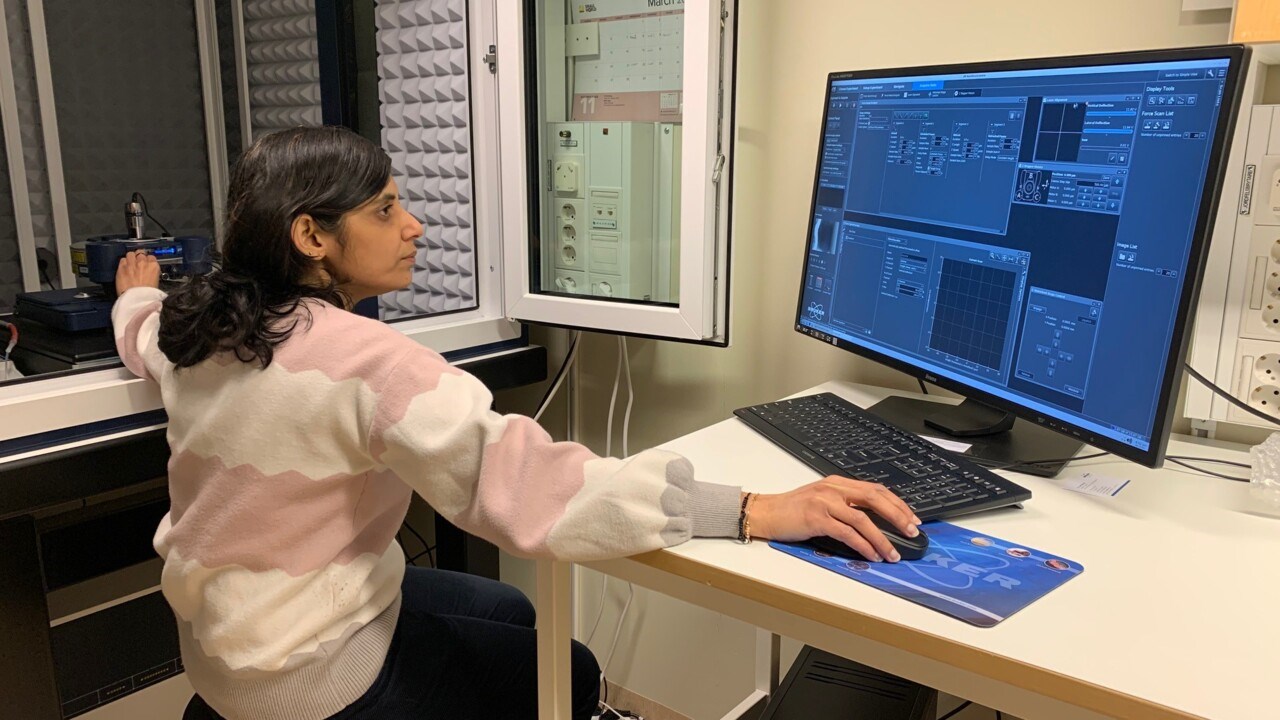
Fozia Bano operating the atomic force microscope.
ImageChloé Jacqueta very attractive technique in both biology and material science
“With an atomic force microscope, researchers can get high quality 3D topographic images on a nanometer scale”, says Fouzia Bano, senior research engineer at the Department of Clinical Microbiology at Umeå University. She is working in Marta Bally’s lab, whose research group is affiliated with both Wallenberg Centre for Molecular Medicine (WCMM) and Umeå Centre for Microbial Research (UCMR).
Atomic Force Microscopy (AFM) is one of the services – and the newest microscope – provided at Biochemical Imaging Centre Umeå (BICU), the research infrastructure at Umeå University. Together with UCEM, BICU forms a node in The National Microscopy Infrastructure (NMI) in Sweden.
Multipurpose technique
AFM can be used to scan various surfaces ranging from metal, glass, and surfaces coated with biomolecules to live cells. It can be used to extract mechanical information such as adhesion, stiffness, and deformation. In addition, AFM can also precisely measure the bond strength and binding affinities of biomolecular interactions.
“AFM is a multipurpose technique. One can use it to image the topography of molecules, quantify the biomolecular interactions, and manipulate the molecules on surfaces. Currently, I am extensively using it to probe biomolecular interactions. For example, using AFM in force spectroscopy mode, I am determining how strong the virus surface proteins bind to their respective receptors on the cell surface.”, says Fouzia Bano.
The working principle of AFM is very simple. It is based on “feeling” the surface by a cantilever with a sharp tip on its free end to scan over the surface, equivalent to the tactile reading of braille.
“The main difference with other imaging microscopic techniques such as electron microscope and confocal is the ability of AFM to provide information about the morphology, assembly, and height, as well as mechanics of samples, all at high spatial resolution and without the requirement of sample labelling. Moreover, the sample preparation is comparatively simple and the possibility of operating AFM in liquid and in various imaging modes make it a very attractive technique in both biology and material science,” says Fouzia Bano.
Studies cell virus entry
Her own current research focuses on unravelling the role of cell surface carbohydrates, specifically glycosaminoglycans (GAGs), in virus entry. Sulphated GAGs such as heparan sulphate is known to play a critical role in modulating the dynamics, such as binding kinetics and diffusion, of many viruses (for example human papillomavirus, herpes simplex virus type 1, and adenoviruses) at the cell surface.
“Using AFM-based single molecule force spectroscopy in combination with other advanced biophysical methods, I am quantifying the binding affinities and the number of contacts for GAG-virus interactions. Besides this, I also use AFM in imaging mode to resolve the self-assembly process of proteins on lipids bilayers”, says Fouzia Bano.
