"False"
Skip to content
printicon
Main menu hidden.

Early development of the nervous system

Our main research focus is to understand molecular mechanisms that regulate the development and function of the nervous system, with focus on the brain and sensory systems.

Head of project

Current projects

At present my research group is interested in four major neural focused projects, and a fifth parallel cancer related project, using a range if model systems.

1) Non-visual opsin related projects:

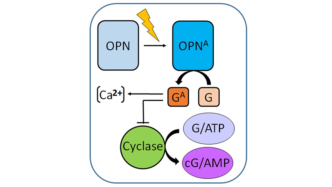
Opsins are G-coupled receptors that detect light by transforming the energy of a photon into a cellular response. Non-visual opsins, not directly involved in visual image formation, are also expressed outside the retina. We are interested to unravel their function in the nervous system, in particular the brain, olfactory and non-retinal eye structures.

Schematic representation of activated OPN (OPNA) signaling.

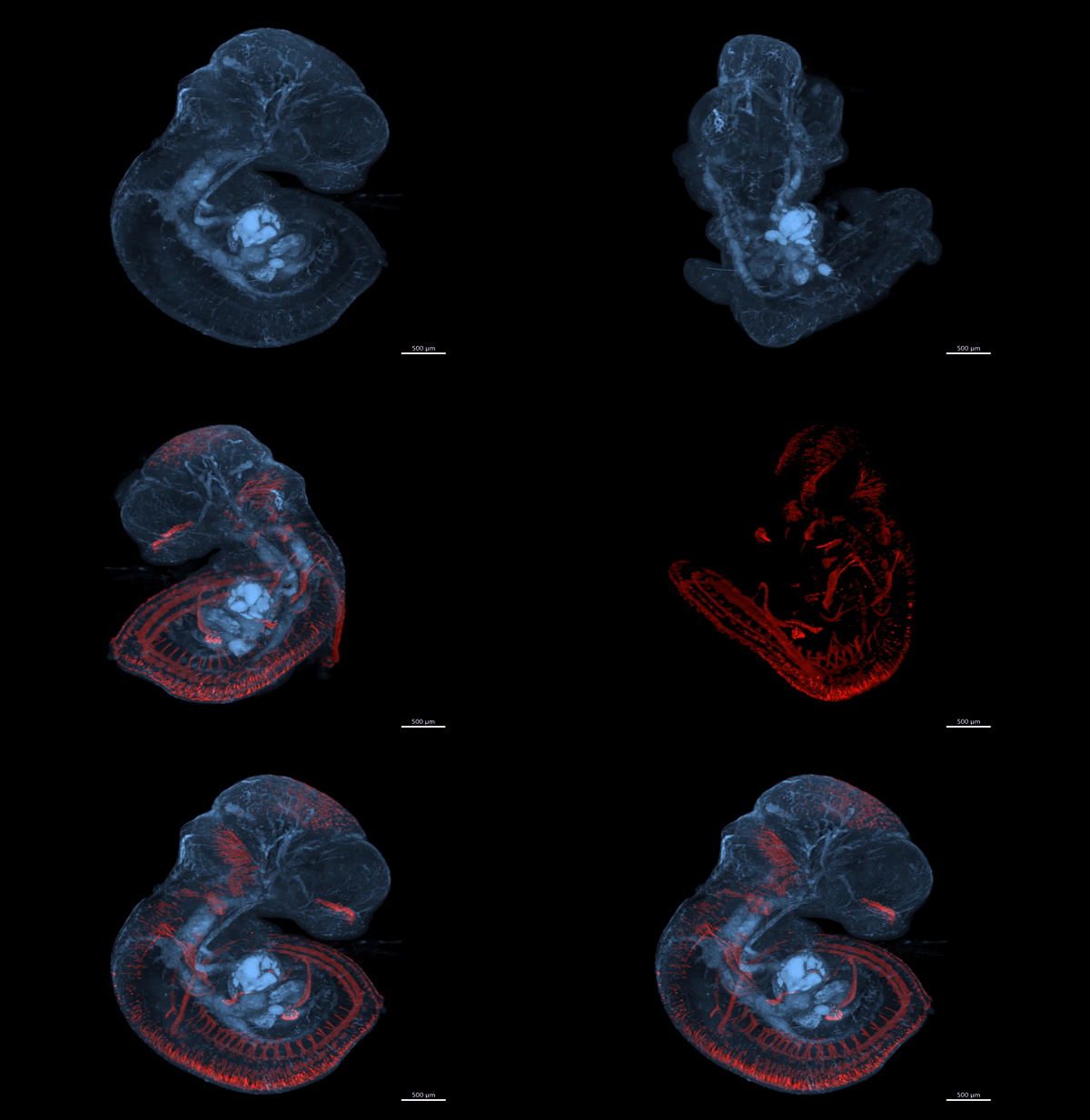
Our recent results1 show a broad but distinct expression of Opsin 3 in the nervous system during early developmental stages. We are now exploring the role of Opsin 3 for the development and function of restricted brain and sensory systems.

1) Davies, W.I.L., Sghari, S., Upton, B.A., Nord, C., Hahn, M., Ahlgren, U., Lang, R.A. and Gunhaga, L. Distinct Opsin 3 (Opn3) expression in the developing nervous system during mammalian embryogenesis.
eNeuro, 15 (8), 0141-21.2021 1–18. (2021).  doi: 10.1523/ENEURO.0141-21.2021

Video S1 - E10.5 Opn3-eGFP Low resolution GIF (002).gif

Movie: Wayne Davies

Movie of 3D imaging of a whole E10.5 embryo showing Opn3-eGFP immunodetection (red) against a background of auto-fluorescing anatomical structures (blue). Movie from Davies et al., eNeuro, 2021.

More results from my lab, done in collaboration with Prof. Richard Lang, Cincinnati, USA, can be found at:  https://scienceoflightcenter.org/basic-science/

2+3) Eye related projects:

The iris in the eye is a sphincter muscle that regulates the size of the pupil, and thereby regulates the amount of incoming light in the eye. Traditionally, it was thought that pupil constriction was solely a brain-mediated reflex, driven by information from the rods and cones in the eye. Now, we and others have shown that light regulated mechanisms directly in the iris is responsible for a local pupillary light reflex. We are expanding our knowledge regarding how the local iris pupillary light reflex is regulated.

The lens and the retina are two important structures within the eye. One interesting aspect of the lens is that stem cells in the lens epithelium proliferate throughout life and give rise to new mature lens fibre cells. The light- and color- sensory cells are found in the retina. We are identifying the molecular actions of different signaling molecules regulating the early generation of lens fiber cells and retina cells, and how these two structures affect the development of each other.

Specific projects related to eye diseases, such as cataract, anolphthalmia and glaucoma are also ongoing.

4) Olfactory related projects:

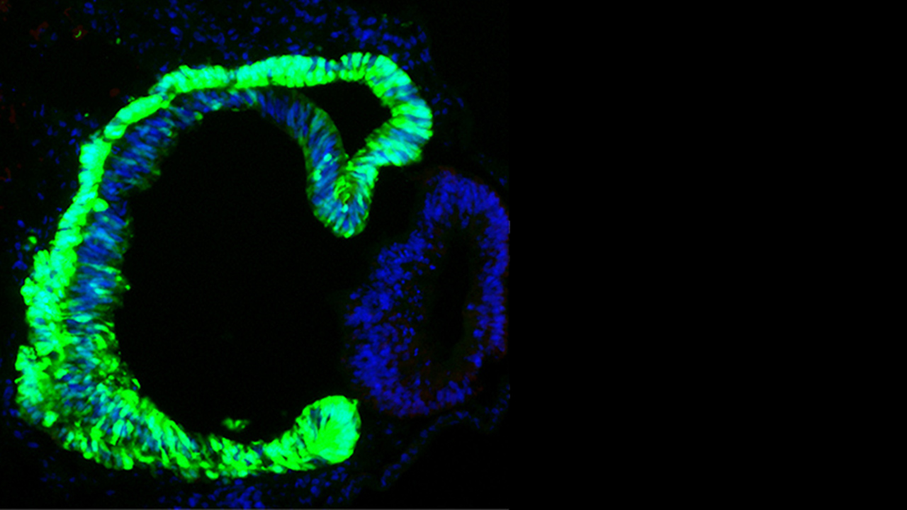
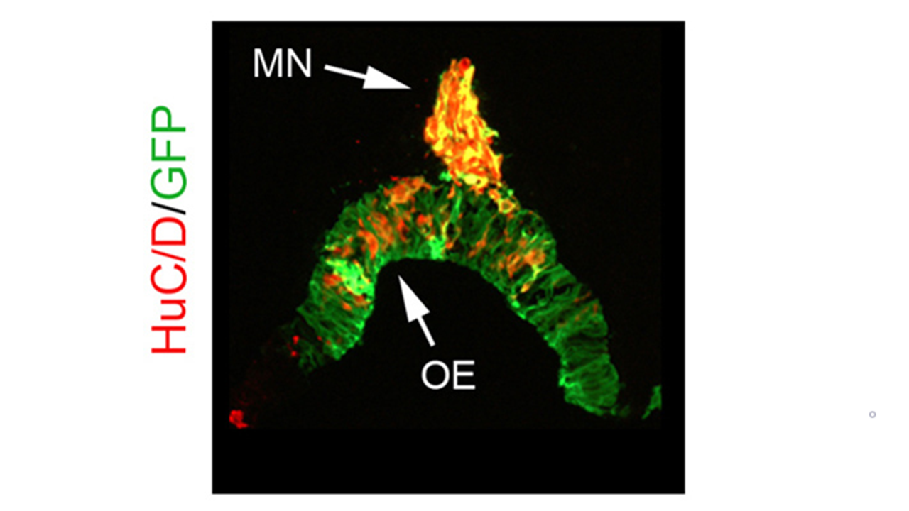
In the adult mammalian head region there are three regions where neurogenesis normally occurs; in which the olfactory epithelium (giving rise to olfactory receptor neurons) is one of the regions. Currently there is a lack of knowledge of the combination and sequence of molecular signals necessary to induce endogenous precursors to efficiently and precisely proliferate and differentiate into appropriate types of neurons within these regions. Using both gain and loss of function approaches in the olfactory epithelium, we aim to unravel the molecular mechanisms regulating the progression from progenitor cells to differentiated neurons.

Moreover, the first post-mitotic neurons in the olfactory epithelium will leave the epithelium by an EMT-like process, including delamination*, and migrate towards the forebrain. We want to define molecular mechanisms that regulate this event, and what function the migratory olfactory neurons have.

* Delamination is a biological normal process where the basal membrane is degraded to facilitate migration of cells. The delamination process is often reactivated in cancer cells, which results in spreading of cancer cells in the body and formation of secondary tumors.

5) Cancer related projects:

We have recently established a delamination/metastatic assay, the CAM-Delam assay, to visualize, score and quantify the ability of cancer cells to degrade the basal lamina (delamination) and migrate. This model is now used in gain-and-loss-of function experiments to determine molecular mechanisms regulating EMT related processes like delamination, invasion and micro-metastasis formation. We are using a range of different human cancer cell lines, such as glioblastoma, lung, colon, prostate and breast cancer cell lines, as well as human tumor samples.

Interested?

If you are interested to work with us as a graduate student (exam-work), Ph.D-student Post-Doc or as a 1st Research Engineer please contact me, Lena Gunhaga at lena.gunhaga@umu.se.

Research Funding:
Our research is / has been funded by, among others:

The Swedish Research Council, Kempestiftelserna, Faculty of Medicine at Umeå University, Ögonfonden, Stiftelsen Kronprinsessan Margaretas arbetsnämnd för synskadade, Strategic Research Area Neuroscience (StratNeuro), Åhlens stiftelse, Märta Lundqvist stiftelse, Cancer Foundation, Cancer Research Foundation Norrland.


Latest update: 2022-10-12