

NEWS Disi Lin has developed new machine learning methods in her doctoral research that incorporate structural information from medical images while also quantifying the certainty of their predictions. "These methods can deliver more reliable analyses and have been tested to identify brain changes linked to Alzheimer’s disease," says Disi Lin, who is now presenting her doctoral thesis for public defence.
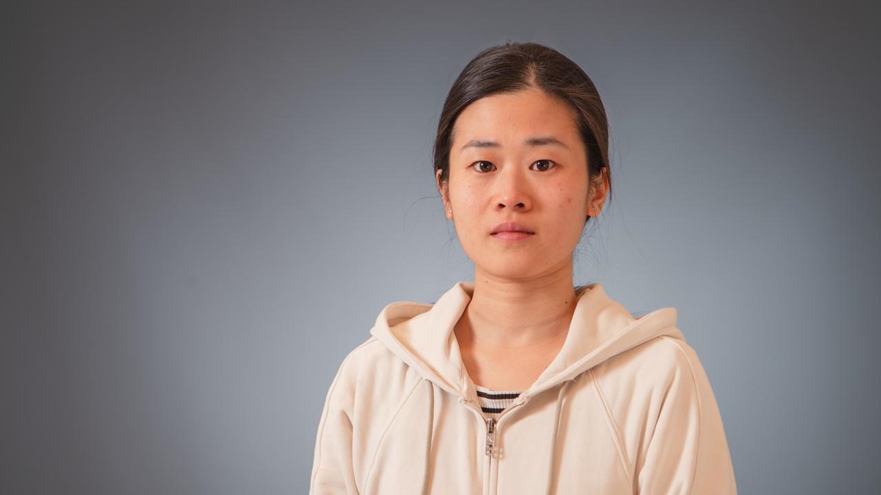
Using advanced mathematical and statistical methods, doctoral student Disi Lin has developed machine learning models that incorporate existing knowledge about human anatomy and can indicate how certain, or uncertain, their predictions are.
ImageHans KarlssonMachine learning enables computers to learn from data and then use that knowledge to make predictions or decisions. Within healthcare, machine learning is used to support tasks such as disease diagnosis, predicting disease progression, treatment planning, and patient monitoring.
"Applying machine learning to medical images is particularly challenging because datasets are often limited, while the models must be both reliable and interpretable," says Disi Lin, doctoral student at the Department of computing Science, Umeå University.
In her doctoral research, Lin has addressed these challenges by integrating prior knowledge into machine learning models. This includes using mathematical morphology, a technique that can be used to encourage the learned patterns to form meaningful structures.
One of the application areas explored is the classification of Alzheimer’s disease.
"Alzheimer’s disease is associated with gradual structural changes in the brain, including reduced volume in brain regions that are important for memory and cognition," says Disi Lin.
Her research combines classical image mathematics with modern machine learning, resulting in models that can identify connected and anatomically meaningful regions more effectively than previous approaches.
"This makes it easier to relate the resulting patterns to disease-related changes and increases confidence in the assessment," says Disi Lin.
As part of her thesis, she has developed methods that take into account the fact that neighboring areas in an MRI scan often consist of the same type of tissue. This reduces the impact of random variations and makes the results more stable and reliable.
"I have also developed methods that not only provide a prediction but also indicate how confident the model is in its prediction. This allows clinicians to see which parts of the analysis are based on robust calculations and which are more uncertain," says Disi Lin.
Disi Lin has incorporated prior knowledge about image structure into machine learning models to help computers identify connected and spatially coherent regions. Rather than treating pixels independently, her AI models can focus on the most relevant areas of the brain, making the results more coherent, interpretable, and medically meaningful.
“These methods can also show how certain the predictions are. This is important in healthcare because obtaining a result is not enough; clinicians also need to know how confident that result is,” says Disi Lin.
Please read the full thesis here: Structure-Aware Machine Learning for Medical Image Analysis.
Disi Lin will defend her doctoral thesis on Friday the 4th of September. Supervisor: Tommy Löfstedt, Docent and Associate professor at the Department of Computing Science.