About the scientific publication
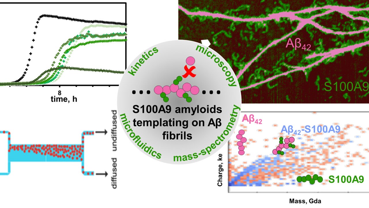
Templating S100A9 amyloids on Aβ fibrillar surfaces revealed by charge detection mass spectrometry, microscopy, kinetic and microfluidic analyses
Jonathan Pansieri, Igor A. Iashchishyn, Hussein Fakhouri, Lucija Ostojić, Mantas Malisauskas,a Greta Musteikyte, Vytautas Smirnovas, Matthias M. Schneider, Tom Scheidt, Catherine K. Xu, Georg Meisl, Tuomas P. J. Knowles, Ehud Gazit, Rodolphe Antoine, Ludmilla A. Morozova-Roche
Chemical Science
https://doi.org/10.1039/C9SC05905A