

NEWS Marine microorganisms play a vital role in regulating ecosystems and the climate. Despite their importance, many of them have never been imaged in detail in their natural environment. Through a new project, postdoctoral researcher George Westmeijer is building a high-resolution image library of microbial life in the Baltic Sea.
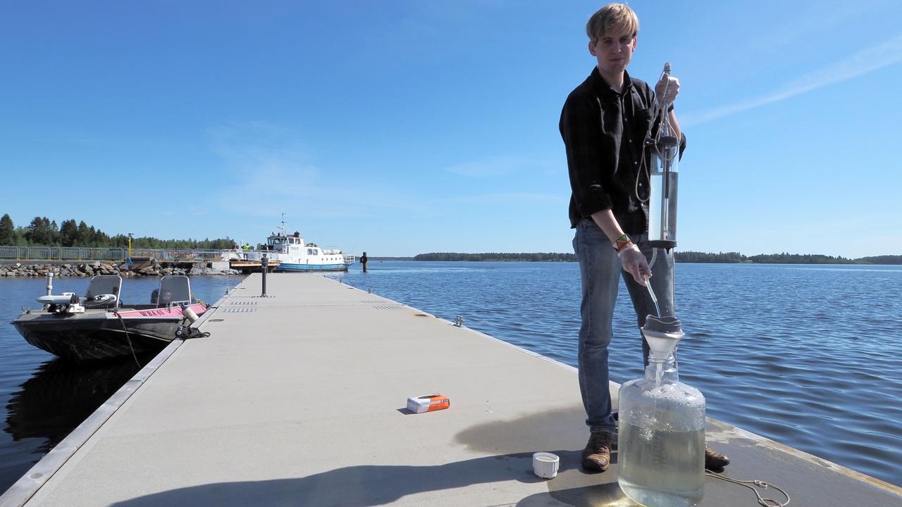
George Westmeijer collecting a water sample at the Umeå Marine Sciences Centre in Norrbyn during the summer. After sampling, the microorganisms are concentrated and examined using light microscopy at the field station before being analysed in greater detail with electron microscopy at Umeå University.
ImageLinda SandbladWe know a lot about the genes of these microbes, but much less about what they actually look like and how they live
"We know a lot about the genes of these microbes, but much less about what they actually look like and how they live. I want to literally see the organisms behind the data and graphs," says George Westmeijer, postdoctoral researcher at the Department of Chemistry at Umeå University.
George Westmeijer is a biologist from the Netherlands who moved to Sweden to pursue his PhD at Linnaeus University in Kalmar. During his PhD studies, he developed a strong interest in microscopy. After moving to Umeå, he came across an advertisement for a postdoctoral position at Umeå University. The project, funded by the Kempe Foundations, felt like a perfect match for his background in ecology and his growing interest in advanced imaging techniques.
The goal of his current project in Umeå is to visualize aquatic microbes – bacteria, archaea, viruses, and their interactions. At Umeå University’s Marine Sciences Centre in Norrbyn, George Westmeijer and his supervisors Linda Sandblad and Johan Wikner have developed a workflow for sampling Baltic Sea water enabling study of microbes at high resolution across seasons and under different environmental conditions.
George Westmeijer chose to focus on the Baltic Sea for both scientific and practical reasons. Its brackish waters host a highly diverse microbial community that changes dramatically with the seasons. The sea’s proximity to the Umeå Marine Sciences Centre, located directly on the coast, also makes year-round sampling possible. During summer, some microbial populations become highly abundant, only to decline to low levels or enter dormancy during the winter as light levels and temperatures are reduced.
One major challenge is that microbes are both small and sparse. Natural seawater contains too few microbes for high-resolution imaging. By concentrating the samples, George Westmeijer increases the number of cells without damaging them.
From sampling to microscopy, the entire process takes about one day.
“I rapidly freeze the samples at very low temperatures, -180 Degrees Celsius. This prevents ice crystals from forming and allows me to study the microbes in a state that is very close to how they exist in nature.
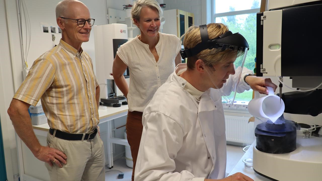
Microscopy is carried out at the Umeå Centre for Electron Microscopy (UCEM). Postdoctoral researcher George Westmeijer prepares samples at minus 180 degrees Celsius. In the photograph, he is pictured with his supervisors Johan Wikner, Professor at the Department of Ecology and Environmental Science, and Linda Sandblad, Associate Professor at the Department of Chemistry.
ImageIngrid SöderberghGeorge Westmeijer then uses advanced cryo-electron tomography (cryo-ET) to create three-dimensional images of microbial cells in the samples. The technique is based on cryo-electron microscopy (cryo-EM), which was awarded the 2017 Nobel Prize in Chemistry.
Cryo-ET combines many images taken from different angles, revealing structures that are impossible to see with conventional microscopy. The image analysis is carried out using software partly developed at the Umeå Centre for Electron Microscopy (UCEM) at Umeå University. For the microscopy part of the project, George is supervised by Linda Sandblad, who is both his principal supervisor and the Director of UCEM.
The resulting image library will have a level of detail that makes it possible to study biological structures such as membrane vesicles, and also interactions between cells.
“Microbes do not live in isolation. We often find different species right next to each other, and we are interested in understanding what kinds of relationships they have and how they influence one another."
With an ever-growing library of microbial images now in place, George Westmeijer is taking the next step. By combining cryo-EM with metagenomics, cell sorting, single-cell sequencing and proteomics, he hopes to identify species, estimate biomass and track developmental stages. The approach could also reveal microbial partnerships between species that are regularly found together.
“Identification is the big challenge ahead,” says George Westmeijer. “But if we succeed, we can start building three‑dimensional references of common marine microbes and explore how their morphology changes in response to factors like rising water temperatures.”
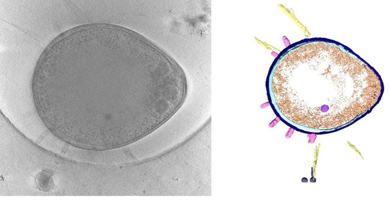
Left: A black-and-white slice from an electron tomogram of an aquatic bacteria. Right: A segmented reconstruction highlighting key cellular structures, including the double membrane, stalks, and ribosomes. The coloured 3D model of the cell was created in Dragonfly.
ImageGeorge WestmeijerIn the long term, the image library could help reconstruct microbial food webs and improve our understanding of how marine ecosystems respond to climate change and human impact. For George Westmeijer, the motivation is also personal:
“For the first time, I truly get to see the organisms I have studied throughout my scientific career. It is fascinating, and possibly the beginning of a new way to understand the most fundamental life in the sea.”
George Westmeijer has a KBC postdoctoral fellowship funded by the Kempe Foundation. The project is interdisciplinary, spanning chemistry and ecology.
