Research project Scar-formation and degeneration of the cornea in the eye and of muscle tendons often lead to deteriorated vision and decreased mobility, respectively. The purpose of this project is to unravel the pathophysiological mechanisms behind such processes.
Collagen degradation and fiber malalignment, as well as apoptosis, angiogenesis, and pain, characterize several pathological conditions of collagen-rich tissues, but the underlying mechanisms are poorly understood. We aim to define intra- and intercellular signaling pathways that regulate the functional states of the collagen-synthesizing cells in muscle tendons and the cornea. One of our hypotheses is that locally produced neuromodulators contribute to excessive scaring by inhibiting apoptosis. The goal is to identify key processes that can be targeted in future intervention techniques preventing or reversing scar-formation.

Main external funding:
2018-21 | National Swedish Research Council: 4,8 MSEK
2014-17 | National Swedish Research Council: 5,044 MSEK
2010-12 | National Swedish Research Council: 2,1 MSEK
2011-15 | Swedish Society of Medicine: 1,2 MSEK
2016-17 | Cronqvists Foundation: 0,5 MSEK
2011-13 | Cronqvists Foundation: 0,4 MSEK
The overarching purpose of this project is to unravel the pathophysiological mechanisms that underlie scar-formation and degeneration in collagen-rich tissues, in order to facilitate development of new intervention techniques that can mitigate identified deleterious processes in such conditions.
More specifically we aim to define intra- and intercellular signaling pathways that regulate the functional states of the collagen-synthesizing fibroblasts of the cornea (keratocytes) and muscle tendons (tenocytes). To achieve this, our studies address (1) the roles of neuropetides and other classically neuronal modulators that we have shown to be produced in collagenous tissues, especially under pathological conditions, (2) mechanisms that inhibit fibroblast apoptosis, and (3) the importance of induction of fibroblast senescence.
The functions of tissues rich in collagen, like tendons and the corneal stroma, depend on an intricate regulated turnover of cells and extracellular matrix. Conditions in which this regulation is affected are characterized by collagen degradation and structural fiber malalignment, apoptosis, angiogenesis, and pain. Trauma, infection or degenerative diseases of the cornea can, for instance, result in excessive scaring that leads to impaired vision or blindness. Another example is degenerative tendon disease, tendinosis, which causes chronic pain and decreased mobility. The cellular and molecular mechanisms behind these conditions are poorly understood.
Therefore, these are the principal aims of our research:
A parallel aim of the proposed project is to develop reliable in vitro models that we hope will increase the efficiency of this research field, complement established in vivo models, and reduce the need for animal experiments. We have successfully developed such a model system for tendons and we will now go further by developing an in vitro model system for the cornea.
Potentially, this research has considerable clinical implications. The results might make possible new minimally invasive treatment regimens for tendinosis and excessive corneal scaring. That is, the research might lead to introduction of novel therapies based on topically/locally administered medicaments, thus reducing the need for the radical surgical methods used today.
Depending on sport, up to ~50% of elite athletes suffer from prolonged and severe pain and reduced mobility due to tendinosis, and about 1/20 of people living a sedentary life-style are similarly affected. Therefore, the need for improved treatments is evident. For athletes, tendinosis often leads to the end of a career, and for people with cardiovascular disease tendon pain might prohibit vital exercise – tendinosis is associated with metabolic syndrome.
According to WHO, corneal opacities cause ~5% of all blindness in the world, being the 4th leading cause of vision loss globally. Many patients with excessive corneal scaring, may it be by trauma, infection or degenerative disease, would benefit from new ways of prevention and/or treatment. In the developed world, alternatives to radical corneal surgery would be welcome, whereas in undeveloped countries, where surgery is not even available, help based on topical treatments would have a great impact. Corneal transplant is the only available curative treatment today, but even in many developed countries, it is difficult to get access to this surgery due to lack of donors.
Principal Investigator (PI) for the project is professor Patrik Danielson, Department of Integrative Medical Biology.

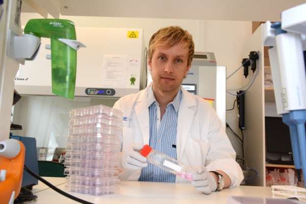
Prof. Patrik Danielson.
PhotoJan Alfredsson